Note: All work composed and submitted by Nicholas Angeles on the respective dates provided. The following work has been pasted from the original documents of the assignment for ease of access:
Draw IgM: Submitted 02/15/26
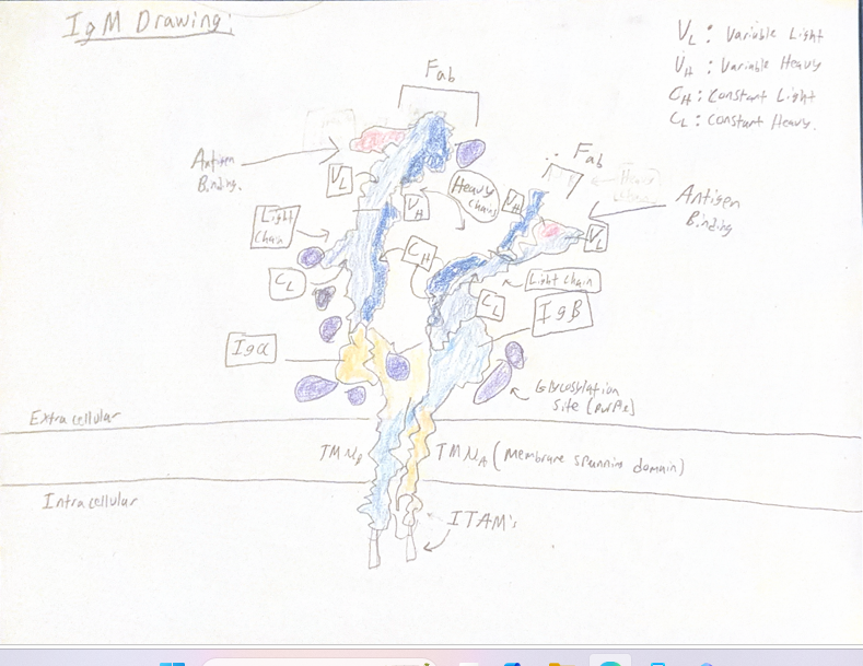
In the figure drawn of IgM in its monomeric, membrane-bound form (mIgM) it contains
a few interesting features that have been illustrated to emphasize the importance of this structure as a BCR, such as Fragment-antigen binding sites (Fab) towards the top of the molecule that consist of variable regions (VH and VL) which can recognize diverse and unique amino acid sequences of antigens. This drawing also shows the different chains that make up the mIgM molecule, such as the light chain, the heavy chain, and the Igɑ and Ig chains. These Igɑ and IgB chains specifically facilitate signal transduction with the use of intracellular ITAMs
indicated at the bottom of the image. This structure differs to that of IgM in its soluble,
pentameric structure in many different ways, such as mIgM’s use of six chains instead of 21 alike soluble IgM as it is membrane bound. Soluble IgM is identified by its iconic pentameric
structure, with five monomeric units that have joined together due to prevalent Cys residues
forming disulfide bonds holding the structure together, whereas the drawn structure mIgM has
only one monomeric unit. This drawn structure also contains immunoreceptor tyrosine-based
activation Motifs (ITAMs) that are utilized by the alpha and beta chains of the structure, whereas soluble IgM has no ITAMs to communicate with its different chains, as it is not membrane bound, and therefore does not initiate signal transduction in a cell directly. The structure of mIgM overall constitutes its role as a B-cell receptor as its properties allow their B-cell to take on many immunological functions critical to the recognition and defense against various antigens.
Find me a mAB!: Submitted 03/08/26
Monoclonal antibodies (mAb’s) are typically characterized by their artificial production and ability to fight various pathogens via their designed antibody features that mimic naturally present antibodies. Monoclonal antibodies can fight various types of pathogens, such as viruses or bacteria, but some in particular are useful for slowing the growth of malignant tumors via their signaling capabilities in cancerous cells/tissues. One fascinating mAb that has growth suppressing properties in cancerous tissue would be a mAb known as Trastuzumab, which specifically targets the function of the Human Epidermal growth factor Receptor 2 (HER2). HER2 is a transmembrane protein responsible for growth in stratified squamous epithelia
systemically, but mainly pertains to breast tissue growth along with limited functions in growth of esophageal and GI tissue alike, but not primarily. HER2 is classified as a Receptor Tyrosine Kinase (RTK) belonging to the HER family, and contains three specific domains of receptor function that mediate growth of the cell. 1) First domain is the extracellular domain that are involved with ligand-dependent/independent dimerization, 2) second domain is the transmembrane domain which will connect the extracellular components to the cytosol of the cell, allowing for growth signal cascades, and 3) the third domain is the C-terminal cytoplasmic domain which will allow for tyrosine kinase activity of the receptor to induce response in the cell.
Of course because HER2 is a growth factor receptor, it is understood that overexpression can cause cancer in its host cells/tissue by causing large amounts of growth signals to cascade in the cell, which leads to over proliferation of epithelial cells and uncontrolled growth. (Maadi et al.) Understanding the structure and respective functions of regions of the HER2 receptor allows strategies to be developed to target its function. It is said that HER2 overexpression is prevalent
in about 20-30% of breast cancer patients, so mediation for this receptor needed to be investigated, which is why Trastuzumab was produced and specifically designed to repress the function of the HER2 receptors that have been overexpressed, ultimately limiting the overgrowth of breast tissue. (Mitri et al.)
Trastuzumab is a class IgG antibody, specifically IgG1 Kappa antibody. IgG antibodies demonstrate the following structure shown below:
Figure 1. IgG Antibody Structure; Soluble, Non-Membrane Bound:
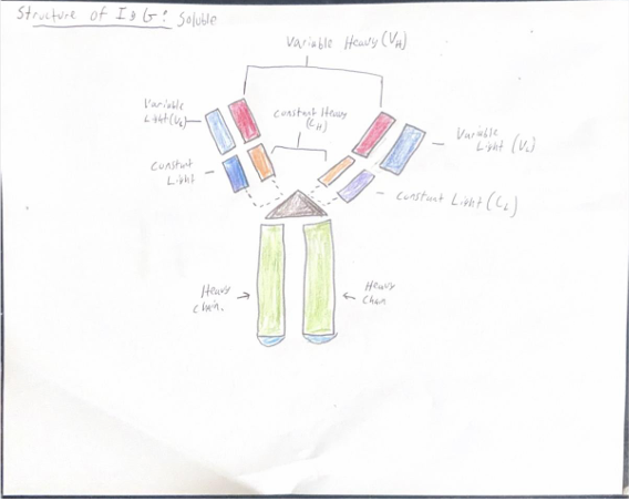
In Figure 1 it can be seen that IgG will contain its classical light and heavy chains, those being variable or constant, along with two heavy chains acting as the “body” of the antibody. The variable region specifically will act as the (Fab) or Fragment Antigen Binding, which plays a role in binding antigens or other proteins to the antibody. More specifically in the case of trastuzumab, the antibody physically binds to and recognizes HER2 directly on the surface of the cell, which causes inhibition of HER2 mediated signaling to promote the growth of the cell. This will firstly inhibit HER2 heterodimerization, which will prevent many different processes in the cancer cell. One process that HER2 allows for in the epithelial cell is a specific MAPK signaling pathway, which allows for growth and proliferation of the cell. When Trastuzumab is bound to HER2, however, it inhibits the initiation of this signaling pathway, which of course prevents the cells growth and proliferation. This is a good mediation technique by Trastuzumab because it blocks a key pathway that causes malignancy in patients with breast cancer. Another process inhibited by binding of Trastuzumab is the cell cycle, specifically inhibiting signals (by extension of MAPK inhibition) that will cause gene activation for cell division. Another quite interesting and important function of Trastuzumab in slowing growth of cancer cells is by inhibiting the actual cleavage of HER2 receptors into their more active, p95HER2 forms which are known to be constitutively active and have a more so called “aggressive” effect in cell proliferation. With Trastuzumab blocking this cleavage, it reduces oncogenic functions of HER2 and overall works to reduce the mitotic index of cancerous cells affected by HER2
overexpression. (Maadi et al.)
Given this information, it can be seen that Trastuzumab is a very useful monoclonal
antibody in its mediation techniques to breast cancer playing a role in regulation of the HER2 receptor, inhibiting many functional pathways to proliferation of affected cancer cells by the HER2 receptor, and overall presenting hopeful solutions to breast cancer by mediating the 20-30% of breast cancer that is considered HER2 positive.
References:
Maadi, H., Soheilifar, M. H., Choi, W.-S., Moshtaghian, A., & Wang, Z. (2021, July 15).
Trastuzumab mechanism of action; 20 years of research to unravel a dilemma. Cancers.
https://pmc.ncbi.nlm.nih.gov/articles/PMC8303665/
Mitri, Zahi, et al. “The HER2 Receptor in Breast Cancer: Pathophysiology, Clinical Use,
and New Advances in Therapy.” Chemotherapy Research and Practice, U.S. National Library of
Medicine, 2012, pmc.ncbi.nlm.nih.gov/articles/PMC3539433/ Accessed 08 Mar. 2026.
End of Term Reflection: New
When reflecting on this course and the many different components to its’ structure, I can confidently say that there were many concepts that have overlapped with my other studies which I have honestly quite enjoyed. To pinpoint all of those overlaps would be quite difficult, however one notable topic that I have learned this semester that tied into my biological and biochemistry coursework would be the various immunological pathways that I have learned and studied this semester, which range from learning about pathways of specific receptors to complement pathways to responses of lymphocytes to pathogen. The immunological pathways that I’ve studied this semester have proven relevance to my studies of biochemical pathways intracellularly when discussing general cellular conditions in my biochemistry lecture. Specifically, when I was studying biochemical pathways involved in cellular respiration in biochemistry, I learned how cells in general have similar cascading techniques such as phosphorylation and dephosphorylation in order to relay physiological responses of the glycolytic or TCA pathways to generate cellular energy. I’ve come to find that the same is true of immunological considerations involving response pathways, where some receptors when bound to extracellular ligand would cause a phosphorylation cascade intracellularly to induce responses in immune cells like upregulation of transcription factors to compose adequate and sometimes directly combative responses to environmental stimuli and stresses (like pathogen). This class has helped me to understand a larger picture of the concepts that I have already been familiar with, and to be more open-minded about general cellular mechanisms that I may study or have studied in the past, with intent to apply it directly to specific cases (such as immunological considerations) and even clinical considerations, which I have found the most interesting cases to apply these foundations to. This class has made me question how biochemistry specifics tie into the topics we discussed in an immunological context as well, such as how lymphocytes regulate ATP synthesis different than other cells based on their needs to respond rapidly and continuously in some cases. I would say that this class overall has helped me to think more critically about biological systems and to use that critical thinking to ask great questions about the systems that I will learn in the near future, to not only feed my fascination but also to help me gain a better understanding in the course content to come next in later semesters, graduate school and beyond.