Scientific Literature Essay
Three major organelles that are thought to have a role in cell death are the chloroplast and pyrenoid, the mitochondria, and the nucleus. The pyrenoids are in the chloroplast of different algae or in hornwort plants. The main function of pyrenoids is to act as the center of carbon dioxide fixation by maintaining a carbon dioxide rich environment around the enzyme ribulose biphosphate carboxylase (Wikipedia, 2021a). Ribulose biphosphate carboxylase or RuBisCO is one of the enzymes that plays a large role in photosynthesis. Chloroplast role in programmed cell death (PCD) is critical when light is involved. In chloroplast, reactive oxygen species (ROS) are important to the programmed cell death (Aken and Breusegem, 2015). This ROS can be found in chloroplasts due to the electron transport chain. That is why mitochondria also play a role in programmed cell death because of their electron transport chain (Aken and Breusegem, 2015). The mitochondria also have reactive oxygen species which are important to programmed cell death. The nucleus role in apoptosis differs from mitochondria and chloroplast. When apoptosis occurs, the chromatin condenses resulting in the nucleus condensing as well. These condensed chromatins can only be by the nuclease, Caspase-activated DNase (CDN). Activation of CDN creates fragments of many base pairs. This DNA fragmentation also causes more cells to be stopped at thesubG1 location. These subG1 cells are apoptotic cells, but this includes all dead cells not just apoptotic cells. Nuclear membrane blebbing is another role that the nucleus plays in apoptosis. This is when during apoptosis, the membranes bulge out of the cell eventually becoming apoptotic bodies that are not part of the old cell.
The algae that we are evaluating are Chlorella, Pandorina, Rhodochorton, and Volvox. The algae Chlorella is green algae, that is unicellular (Wikipedia, 2021b). It is found in freshwater and it does contain chloroplasts. With it being a green alga, it also does have pyrenoids in its chloroplasts. The algae Pandorina is also a green alga which means in its chloroplasts, pyrenoids will also reside to contribute to the photosynthetic process. These Pandorina are multicellular being composed of either 8, 16, or even 32 cells (Wikipedia, 2021c). While Chlorella and Pandorina are both green algae, both reside in freshwater, but one is unicellular, and one is unicellular and colonial. Rhodochorton is another alga we are evaluating that is of the red algae unlike the previous two green algae. Unlike the other algae we have looked at, Rhodochorton resides mainly in marine environments, but does sometimes reside in freshwater (Wikipedia, 2020). Rhodochorton does have chloroplasts, but it does not have the pyrenoids like Chlorella or Pandorina. It is a unicellular organism that usually brakes off into branches or threads. The final algae we are going to evaluate is Volvox. This is a colonial unicellular organism, but its colonies are much larger than those of Pandorina. The colonies of Volvox can be up to 50,000 cells that live in freshwater environments (Wikipedia, 2021d). Volvox just like the other green algae, have a chloroplast that does contain pyrenoids.
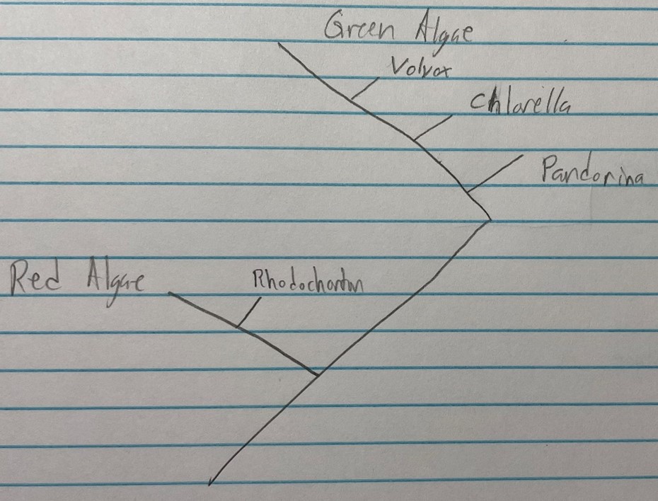
The evolutionary history of green and red algae differs quite significantly. Green algae are more closely related to plants than red algae. If plants were also on the phylogenetic tree below, that would be shown as the green algae branch would be closer to the plant branch than the red algae branch. In this evaluation, we looked at three different green algae: Volvox, Chlorella, and Pandorina. In comparison, we only looked at one red algae: Rhodochorton. Green algae are more commonly found in freshwater environments and red algae are more likely to be found in marine environments. Overall, from the phylogenetic tree below, we see that we are looking at three green algae and one red alga and that while both are classified as algae, they are not as closely related as some may think.
In the scientific paper Experimental taphonomy of organelles and the fossil record of early eukaryote evolution, they began the experiment with four algae species. These four species were Volvox aureus, Pandorina morum, Chlorella, and Rhodochorton and each was killed in a 300 mM of β-mercaptoethanol (BME) for one day. Once this day had passed, the excess BME was removed, and the algae were rinsed with each of their corresponding water types. These samples of algae were then collected to be examined in the experiment. The presence of oxygen is known as oxic and the absence of oxygen is known as anoxic. In the experiment they used thirty-two 1mL test tubes among each of the algae and separated them into the oxic and anoxic groups. In the oxic group they left the lids off and kept the tubes hydrated with water on top. In the anoxic group, the lids were left on the test tubes and they were sealed. To prevent temperature changes from becoming an unwanted contributing factor to the experiment, the test tubes were all placed in an incubator where the environment was left at 17.5°C. On top of that, these test tubes were sampled every two to four days for a period of a month to ensure that the temperature was being maintained. From this experiment, they noticed that the anoxic conditions did show slower decay rates (Carlisle, Jobbins, Pankhania, Cunningham, Donoghue, 2021). On the other hand, they did not notice any effects from the oxic or anoxic conditions on the decay patterns (Carlisle, Jobbins, Pankhania, Cunningham, Donoghue, 2021).
In their experiment, the authors chow multiple features of the cell. These features include if the nucleus was visible, if the chloroplasts had thinned, if the pyrenoids were visible, if the cell had collapsed, if the chloroplast had holes, and if the chloroplast had collapsed (Carlisle, Jobbins, Pankhania, Cunningham, Donoghue, 2021). First, we will look at the Volvox aureus which was examined for a period of a month. The nuclei were still present and visible throughout the month. If they were to disappear, the nuclei were more likely to disappear before the chloroplasts as seen in Figure 2. Chloroplasts however thinned, deformed, and perforated before disappearing in the images. We also saw that even with pyrenoids being the most likely to decay, they left behind a starch grain ring where they were in Figure 2. In the Pandorina morum, we see that they collapsed right after they had been placed in the incubator which would cause the cells to lose y-shaped junctions (Carlisle, Jobbins, Pankhania, Cunningham, Donoghue, 2021). From looking at Figure 2, we can also see that after the month in the incubator the nuclei were still visible. The only other change observable was that the number of nuclei had decreased as the weeks went on. When looking at the chloroplasts in the Pandorina morum, we can see that they had thinned and had holes throughout after the month. These indications lead me to believe that with a couple more weeks, the chloroplasts would soon disappear. The pyrenoids decayed leaving behind starch grain rings just as they had in the Volvox aureus. In the Chlorella sample, the nuclei were visible throughout the experiment in some of the cells, while in others they were not visible. The pyrenoids of the Chlorella disappeared immediately and the chloroplast had holes, thinning, and less chloroplasts were visible junctions (Carlisle, Jobbins, Pankhania, Cunningham, Donoghue, 2021). In Figure 3, we can also see the Rhodochorton which had visible nuclei throughout the month. The chloroplasts of the Rhodochorton decayed immediately resulting in holes, deforming, and thinning. In conclusion, the Volvox aureus showed a visible nucleus. The chloroplasts thinning and holes which would result in the deforming of them. The pyrenoids were not visible and had completely disappeared from the chloroplast. The Pandorina morum cells had a visible nucleus throughout the month. They also had chloroplasts that had thinned and developed holes. The Pandorina morum’s pyrenoids were not visible as they decayed rather quickly. The Chlorella cells had visible nuclei with no signs of decaying. The chloroplasts thinned and had holes and the pyrenoids of the Chlorella cells had decayed immediately. The final algae Rhodochorton had visible nuclei in the colony. The chloroplasts however had thinned and had holes because they began decaying immediately when in the incubator.
Figure 4 shows fossil organelles that were analyzed and examined to determine if features of plant cells were visible in the fossils. In the Zelkova leaf fossil, grana stacks and starch grains were seen inside of a chloroplast found in the cell wall (Carlisle, Jobbins, Pankhania, Cunningham, Donoghue, 2021). We can also see that in the Royal fern stem segments, nuclei are visible within each cell. Image C from Figure 4 also shows signs of nuclei, but no definitive data on whether or not nuclei are identifiable has been found (Carlisle, Jobbins, Pankhania, Cunningham, Donoghue, 2021). The idea that chloroplasts and nuclei could remain intact in the fossils of some plants does support the findings from the experiment that the nuclei and chloroplasts are the most decay resistant in the algae that analyzed and examined in the experiment.
Aken, O. V., and Breusegem, F. V. (2015). Licensed to Kill Chloroplasts, Mitochondria, and Cell Death. Trends in Plant Science 20, 754-766.
Carlisle, E., Jobbins, M., Pankhania, V., Cunningham, J., Donoghue, P. (2021). Experimental taphonomy of organelles and the fossil record of early eukaryotic evolution. Science Advances 7, no. 5.
Nuclear condensation DNA fragmentation and membrane disruption during apoptosis. Abcam, https://www.abcam.com/kits/nuclear-condensation-dna-fragmentation-and-membrane-disruption-during-apoptosis
Wikipedia contributors. (2021a). Pyrenoid, https://en.wikipedia.org/wiki/Pyrenoid
Wikipedia contributors. (2021b). Chlorella, https://en.wikipedia.org/wiki/Chlorella
Wikipedia contributors. (2021c). Pandorina, https://en.wikipedia.org/wiki/Pandorina
Wikipedia contributors. (2020). Rhodochorton, https://en.wikipedia.org/wiki/Rhodochorton
Wikipedia contributors. (2021d). Volvox, https://en.wikipedia.org/wiki/Volvox