Neuroscience
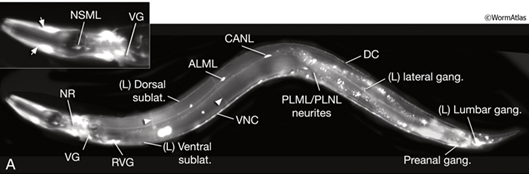
This picture shows the nervous system of C. elegans. Some components of this system are shown such as the nerve ring (NR), retrovesicular ganglion (RVG) and ventral ganglion as well as their directional positions.
C. elegans are not a recent discovery. They were first used in the work of Emile Maupas since the late 1890’s. Then Victor Nigon and Ellis C. Dougherty conducted analyses on the nematodes. However, Sydney Brenner is credited for making C. elegans a prominent research model in the 1960’s and 1970’s. They also rose to fame because C. elegans are the first organisms with multiple cells to be sequenced in 1998. Many findings have been made possible by C. elegans including gene silencing by small RNAs and apoptosis according to Félix and Frézal (2015). C. elegans are used as animal models because they have all the functional attributes of an animal. C. elegans have neurons, skin, stomach, muscles, and other tissues which resembles in shape and role just like the systems in humans. They also use neurotransmitters such as monoamines, acetylcholine, GABA, and glutamate which are found in other organisms like Homo sapiens.
C. elegans have two different unrelated nervous systems. They have a somatic nervous system, which is comprised about 282 neurons. They also have a pharyngeal nervous system which has about 20 neurons. These structures transmit signals through 1 pair of RIP interneurons. According to Altun and Hall (2011), the neurons and processes that occur in the somatic nervous system are situated between the hypodermis and the body muscle wall. The basal lamina separates the hypodermis and muscles from each other. The pharyngeal nervous system rests among the muscles and there is no barrier between them. Nematodes have about 900 gap junctions and 1,500 neuromuscular junctions. They also have close to 6,400 chemical synapse which can be duplicated 75 percent of the time. Chemical synapses happen between presynaptic and postsynaptic cells. Immunochemical staining corroborated that post-synaptic receptors are collected on post-synaptic processes. The neurons of roundworms are categorized into four groups which are motor, sensory, interneurons, and polymodal neurons. Motor neurons are responsible for making synaptic connections for muscle cells. C. elegans move by either crawling on hard surfaces or by a swimming-like motion in liquids, which are carried out by approximately 113 neurons. Sensory neurons are associated with the senses. Roundworms respond to its environment by using aerotaxis, chemotaxis, and thermotaxis. This set of neurons is comprised of about 6 interneurons and 69 motor neurons. Interneurons are responsible for receiving and sending synapses from and to other neurons. They also process information. They are the biggest group of neurons found in roundworms. Polymodal neurons are capable of doing many functions. They can do the functions of motor, sensory, and interneuron in combinations (like interneuron-sensory functions). These neurons are found commonly in the tail of a male (Altun & Hall, 2011).