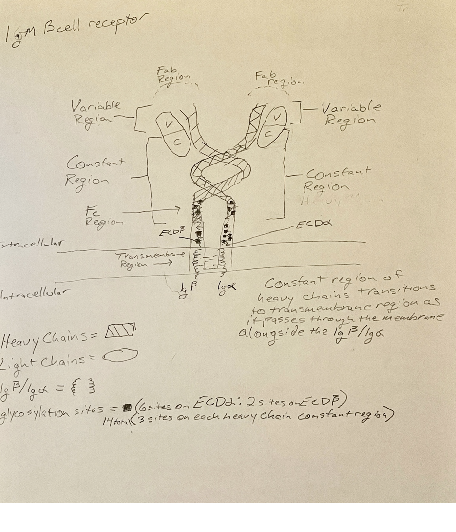
This drawing is a crude representation of the human membrane bound IgM B cell receptor. The complexity of the receptor is difficult to demonstrate. The structure begins in the interior of the membrane as both the Iga/Igb and the heavy chains span the membrane. As the structure develops into the extracellular space the heavy chain constant regions wrap around each other. There are multiple glycosylation sites in the ECDa/ECDb and a few sites in the heavy chain constant regions. As the receptor expands upward, the constant regions stop intertwining and flare out, creating space for the Fab regions. Which consist of the variable regions of both heavy chains and both light chains. Fab stands for fragment antigen binding and is the N- terminal sections of both light and heavy chains. This is the section of the B cell receptor that binds antigen. The membrane bound IgM B cell receptor differs from the soluble IgM B cell in that the soluble version is a pentamer or a hexamer. Their constant regions are similar, but their C terminal structures are very different. Membrane bound IgM B cell receptors have C terminal alpha helices, whereas soluble IgM has C terminal Beta strands. The Beta strands assist with oligomerization, whereas the alpha helices of membrane bound IgM B cell receptors anchor the receptor to the cell surface.
We use cookies to ensure that we give you the best experience on our website. If you continue to use this site we will assume that you are happy with it.Accept

Leave a Reply