
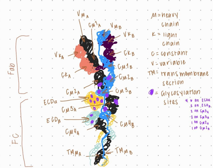
This drawing replicates the cryo-electron microscopy molecule of immunoglobulin M (IgM). In
this model, the different aspects of what makes the immunoglobulin are represented in different
colors to make it easier to see how they interact and intertwine. It contains two kinds of chains; a
heavy chain that is represented by the “μ” micro symbol and a light chain represented by the “κ”
kappa symbol. Along with the two chains, there are constant portions where the immunoglobulin
does not contain antigen-binding domains and variable portions that are variable and responsible
for binding to antigens. There are also 14 sites of glycosylation with their location shown in
purple. The cryo-electron microscopy structure is a much more defined and detailed model of the
IgM. The structure of the cryo-em molecule is more dynamically flexible than the soluble
pentameric IgM. It also goes into great detail to help visualize the native state of the IgM,
whereas the pentameric structure is a model of the IgM in a stable state. With the pentameric
IgM model, you can properly view the different components that make up the structure but it
does not accurately represent how the structure is interacting with the different components of
the IgM itself
Leave a Reply